


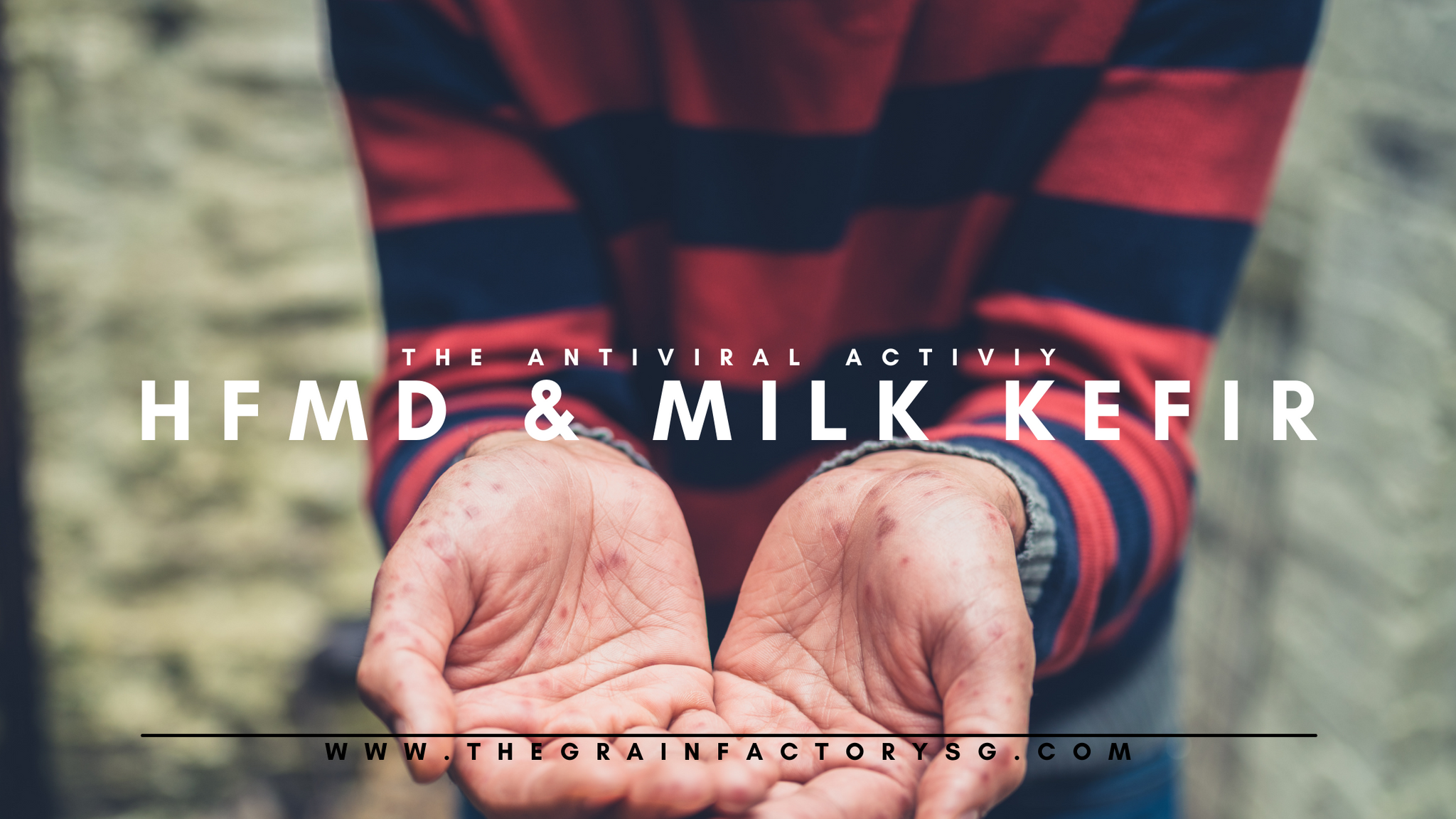
Hand, foot, and mouth disease (HFMD) is an infectious gastrointestinal disease, caused primarily by serotypes of Enterovirus A, most commonly by human enterovirus-A71 (EV-A71) and coxsackievirus-A16 (CV-A16), and mostly affects children.
Coxsackievirus A4 (CV-A4) and A10 (CVA10) have also become increasingly common in recent years, which has coincided with the emergence of more severe cases of HFMD. Herpangina, a more contagious form of HFMD, is mainly caused by a particular strain of group A coxsackievirus, but can also be caused by group B coxsackievirus, echovirus and enterovirus 71.
The incidence of HFMD has also increased over the recent years, making it a very common childhood infection worldwide. While there are no specific treatments for HFMD, a topical oral anesthetic may help relieve the pain of mouth lesions.
These viruses replicate in numbers in our gastrointestinal tract and utilize host gut bacteria to enhance multiplication, pathogenesis, and transmission and thereby lead to development of the disease. The symptoms can include fever, sore throat, rashes and lesions on tongue, gum and inner cheeks.
EV71 linked to more severe case while CV-A4 linked to mild HFMD
The types of virus that cause HFMD differ between mild and severe cases. EV71 was detected at a higher rate in the severe cases, whereas CV-A4 was detected at a higher rate among the mild cases. In 2019, Alexandra et al. found that EV71 can enter the host body via the faecal–oral route and target the gastrointestinal epithelium at the initial stage. This is then followed by the target of the respiratory tract. After that, it gradually infects the host body. Hence in severe cases, neurological and systemic complications can develop rapidly and it can be fatal within 3–5 days in some cases.
Viiral types, viral load and severity of HFMD
We would have thought that the severity of HFMD is solely determined by the type of virus that infects or/and the amount of the virus load.
Shen et al. (2021) has however demonstrated that there was no significant difference in the amount of each virus type between the mild and severe cases. In addition, despite various enterovirus can cause HFMD, the relationship between disease severity and different genotypes of enterovirus remains unestablished till date.
While in another study, a higher amount of the viral load has been found to result in a more severe disease in EVA-71 positive cases only based on throat swabs (Song et al., 2020). The researchers in this study believed that this data is more accurate due to the larger sample size studied as compared to other studies who found no relation with severity and viral load. However due to the assay methods, this above result could not be made certain and more extensive studies are required.
Overall, phylogenetic analysis and viral load tests suggest that neither the genotype of enterovirus nor the amount of virus could be conclusively associated with the increased risk of severe disease.
While there are other factors that may influence the severity of HFMD caused by the same type of enterovirus eg. host immune system, host genetic effects, nutritional and hygiene status, the types of gut microorganisms also plays a significant role and the progression of HFMD.
In this blog, we are focusing on how gut microflora could potentially impact HFMD severity and vice versa.
The prognosis of HFMD is linked to the initial gut microflora status prior to HFMD infection
The initial status of an imbalance of the gut microbiota (termed gut dysbiosis) could mean a higher susceptibility of an upset in the ability of epithelial cells self-renewal, and a dysfunction of the secretion of the mucus layer and tight junctions of intestinal epithelial cells. This can ultimately lead to an intestinal barrier disruption and eventually increase the susceptibility of the individual to immune or inflammatory disorders, including enterovirus and other types of infection.
Our gut microbiome not only works as a physical barrier to prevent pathogens from entering into our systems, they also work in conjunction with the intestinal barrier to orchestrate a defense network that disrupt the invasion of pathogens and maintains the homeostasis and functionality of the gut (Sanz and De, 2009; Odenwald and Turner, 2017; Takiishi et al., 2017; Robinson, 2019).
Furthermore, based on the fact that gut microflora interacts with invading pathogens, the severity of HFMD has also been concluded to be attributed to the enrichment of specific gut microbiota and the gut-derived translocating bacterial surface components that drive inflammation and HFMD viral proliferation (Tan et al., 2022).
The types of gut microbes one have can influence the rate of HFMD viral reproduction and the degree of invasion, contributing either positively or negatively to the illness.
In a similar enrichment analysis, both the gene and species diversity were found to be significantly different between CV-A and EV71 groups. With the discovery of the proportion of severe cases higher in the EV71-positive group than in the CVA-positive group, Bacteroides and Clostridium were detected to be at the highest level in severe EV-positive cases. These results further conclude that the enrichment of these bacteria in a host gut could potentially contribute to the disease severity.
An epidemiological study also found that very few or no mild cases developed into severe cases, whereas severe cases usually progressed quickly, resulting in death in some instances within 1–3 days after the appearance of symptoms]. Mild and severe cases of HFMD might be independent and have different causes and mechanisms.
HFMD mediate changes to host gut microbiome
We knew from earlier discussion about the importance of a healthy balanced gut in maintaining gut barrier function and preventing illness and reducing severity of HFMD.
Many viruses including COVID-19 have cleverly manipulated our gut microbiome environment as one of the strategies to infect deeper and more extensively. Enterovirus and coxsackievirus that cause HFMD and herpangina are no exception. For example, EV71 infection could reduce the expression of goblet cell-derived mucins (Good et al., 2019), which could alter the luminal environment of the gut microbiota and in turn affect the types of gut microbiota present. With EV71 most frequently associated with severe diseases (Shu et al., 2015), the gut microbiota of patients infected with EV71 were more dysbiotic than that of patients with CVA16.
A reduction in the diversity of butyrate-producing bacteria (Faecalibacterium and Bifidobacterium) and an increase in inflammation inducing bacteria (Prevotella and Streptococcus) has been detected in HFMD children. This is an unhealthy shift orchestrated by HFMD invasion.
Prevotella can cause inflammatory reactions and damage the gut barrier (Hofstad et al., 1993; Choi et al., 2011). Streptococcus is one of the most prevalent inhabitants of the respiratory tract (Engholm et al., 2017), skin surface (Ho et al., 2021) and gut (Sokol et al., 2017; Ooi et al., 2018) of adults and children. They are a wide variety of opportunistic pathogens.
Faecalibacterium and Bifidobacterium are producers of short-chain fatty acids and are reported to be capable of regulating intestinal epithelial nutrition, stabilizing immune homeostasis and reinforcing intestinal barrier functions (Hiippala et al., 2018; Routy et al., 2018). Bifidobacteria protect gut mucus against diet-induced microbiota-mediated deterioration, and gut barrier defects (Schroeder et al., 2018). Bifidobacteria have also been identified as health-promoting genera that increases butyrate production and thus contributes to improvements in the gut barrier and metabolic outcomes (Seganfredo et al., 2017). Similarly, Faecalibacterium exhibits an enriched polyphenol-rich dietary pattern, which could improve gut barrier function (Del Bo' et al., 2020).
Based on the above data till date, (1) a well-balanced gut microbiome to resist extensive pathogen invasion and (2) host’s ability to resist microbiota changes due to invading HFMD virus would serve as a potential strategy to prevent or/and reduce the severity of HFMD
Milk kefir support resistance towards HFMD infection due to its anti-viral activity and as a powerful synbiotic food
The microbial community in milk kefir is shown to exhibit antiviral activity against EV71. The researchers maintained that the antiviral outcome was reached through a physical interaction between virus particles and bacteria, which stopped virus admission into the mammalian host cell (Hamida et al., 2021).
Lactobacillus reuteri Protectis, Lactobacillus plantarum and Lactobacillus amylovorus, Lactobacillus acidophilus and Lactobacillus rhamnosus strains are commonly found in milk kefir.
Lactobacillus reuteri Protectis showed substantial activity against EV71. The antiviral activity of the Lactobacillus plantarum and Lactobacillus amylovorus, against enteroviruses has also been demonstrated.. In addition, inhibitory activity against coxsackieviruses was shown by Lactobacillus plantarum, Lactobacillus acidophilus and Lactobacillus rhamnosus strains and their derivatives.
The administration of synbiotic supplements to infected HFMD individuals are found to have lower morbidity, and the gut microbiota showed potential resistance to HFMD-related gut flora dysbiosis which might manifest as a decrease in HFMD susceptibility (Guo et al., 2021).
Synbiotic supplementation was related to an improved protective effect and aided in the enhancement of colonization of beneficial microbes.
In the same study, the results also displayed that children who consumed synbiotics were at lower risk of infection than those who used probiotics alone or prebiotics alone. Although this study did not determine which types of synbiotics was used as there are plenty of food sources that contain prebiotics as well, synbiotics supplementation serves as a potential therapy for HFMD.
Fruits and oats are sources of naturally occurring prebiotic fiber. Milk kefir also contains prebiotics due to kefiran.
Kefiran, a microbial exopolysaccharide (EPS), is found exclusively in milk kefir and has a prebiotic nature which stimulates the growth of healthy microbes in the gastrointestinal tract of the human entity. This therefore achieves a balance of the microbiota in the gut (Salari et al., 2022).
Mix your favorite fruits like banana and avocado with our milk kefir for a more synbiotic boost. And if you are adventurous enough, our organic LACTOats™ (lacto-fermented oats) has so much to offer. It is high in digestibility, packed with nutrients and bursting with prebiotics and probiotics. This makes it an ideal synbiotic food to include in your diet regularly.
References
Shen, C., Xu, Y., Ji, J., Wei, J., Jiang, Y., Yang, Y., Yang, M., Huang, H., Zou, R., Fang, C., Zeng, F., Yang, F., Wang, X., Yuan, J., Li, J., Wang, X., Yang, H., Gong, S., Wang, H., … Liu, Y. (2021). Intestinal microbiota has important effect on severity of hand foot and mouth disease in children. BMC Infectious Diseases, 21(1). https://doi.org/10.1186/s12879-021-06748-7
Atarashi K, Tanoue T, Shima T, Imaoka A, Kuwahara T, Momose Y, Cheng G, Yamasaki S, Saito T, Ohba Y, et al. Induction of colonic regulatory T cells by indigenous Clostridium species. Science. 2011;331(6015):337–41.
Salari, A., Hashemi, M., & Afshari, A. (2022). Functional properties of Kefiran in the medical field and food industry. Current Pharmaceutical Biotechnology, 23(3), 388–395. https://doi.org/10.2174/1389201022666210322121420
Tan, L. T.-H., Letchumanan, V., Law, J. W.-F., Pusparajah, P., Goh, B.-H., Letchuman Ramanathan, G. R., & Lee, L.-H. (2022). IDDF2022-abs-0221 the roles of gut microbiota in hand, foot and mouth disease. Basic Gastroenterology. https://doi.org/10.1136/gutjnl-2022-iddf.70

